Reagent | Product Name | CAT# |
|---|---|---|
Wash Buffers | 10X Phosphate Buffered Saline, PBS Phosphate Buffered Saline-Tween, PBST Tris Buffered Saline, TBS (20X) Tris Buffered Saline-Tween, TBST Distilled Water | |
Blocking Buffers | Bovine Serum Albumin (BSA) Casein (3%, TBS) Casein (3%, PBS) | |
Hybridization Buffers | Saline Sodium Citrate (20X) Denhardt's Solution (50X) Denhardt's Solution (100X) | |
Detergents & Permeabilizers | Triton X-100 Tween-20 (100%) Proteinase K Solution (20mg/mL) Sodium Dodecyl Sulfate (SDS, 20%) NP-40 Lysis Buffer (2X) | |
Fixatives | Paraformaldehyde (4%, PBS) Paraformaldehyde (4%, Phosphate Buffer) Paraformaldehyde (3.7%, Fixative Solution) Paraformaldehyde (4%, PBS with Mg & EGTA) Formalin (10%, Phosphate Buffer) Formalin (10%, Acetic Acid Solution) | |
Probe & Antibody Preparation Buffers | Tris-HCL Buffer (1 M, pH 8.0) EDTA (0.5 M, pH 7.4) Sodium Chloride (1 M) Sodium Acetate (20mM) | |
Substrate Buffers |
Following an in situ hybridization (ISH) protocol allows researchers to detect and localize specific nucleic acid sequences within fixed tissues or cells. ISH is a powerful molecular technique that is essential in both research and diagnostic settings, from gene expression studies to chromosomal mapping. By visualizing RNA or DNA using an in situ method, the ISH protocol reveals spatial patterns of gene expression that are often lost in homogenized assays.
Whether you're performing a fluorescence in situ hybridization (FISH) procedure or using chromogenic detection, success depends on high-quality reagents and optimized protocols for each step, from fixation to signal development.
Here's everything you need to carry out our ISH protocol. In addition to Boston BioProducts' high-quality buffers and reagents, you'll need the following materials and equipment:
The following ISH protocol provides a generalized workflow that can be adapted for both chromogenic and fluorescence-based detection methods. Always tailor parameters like probe concentration, hybridization temperature, and stringency washes to your specific tissue type and target nucleic acid.

Fixation preserves tissue structure and nucleic acid integrity, while permeabilization allows probe access. These steps are critical for achieving specific, localized hybridization signals and minimizing morphological artifacts.
Pre-hybridization conditions the sample and blocks nonspecific binding sites. This helps ensure that only your probe binds to its complementary target, improving signal clarity and reducing background.
This is the core of the ISH protocol-when the labeled probe binds to its complementary DNA or RNA sequence. Precise temperature control and adequate incubation time are essential for specificity and sensitivity.
Washing removes unbound or weakly bound probes and reduces background signal. Adjusting stringency through temperature and salt concentration helps retain specific hybridization while eliminating noise.
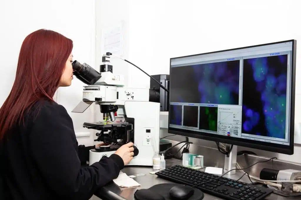
After hybridization and stringency washes, visualization of the probe-target complex is achieved through either chromogenic or fluorescent detection. In a chromogenic ISH protocol, the signal is enzyme-amplified and visible under brightfield microscopy. For fluorescent detection, such as the FISH protocol in situ, directly or indirectly labeled probes allow for sensitive imaging with fluorescence microscopy.
For Chromogenic ISH:
For FISH:

The problem:
Non-specific probe binding, insufficient washes, or inadequate blocking can all contribute to elevated background in your ISH protocol.
What to do:
The problem:
Low probe binding efficiency may result from poor tissue accessibility, probe degradation, or insufficient concentration.
What to do:
The problem:
Patchy or inconsistent staining often results from poor probe distribution or drying during hybridization.
What to do:
The problem:
Off-target binding or tissue damage can cause a misleading signal in your in situ hybridization protocol.
What to do:
For paraffin sections: Ensure sections are fully deparaffinized and rehydrated before fixation. Residual wax inhibits probe binding.
Boston BioProducts offers a comprehensive selection of high-quality reagents specifically formulated for ISH protocols. From fixation to final imaging, our buffers are designed for consistency, low background, and compatibility with both chromogenic and fluorescence-based detection systems.
Need a specialized formulation? We also provide custom buffer services to support unique tissue types, detection chemistries, or workflow needs. Whether you're developing a FISH protocol in situ or troubleshooting background in chromogenic ISH, our products-and our team-are here to help you optimize every step.
Fix samples in 4% paraformaldehyde (PFA) for 15-30 minutes at room temperature.
Wash in PBS to remove residual fixative.
Permeabilize tissue using 0.1% Triton X-100 or treat with Proteinase K for RNA targets.
Formulate your pre-hybridization buffer. Here's a recipe you can use for 100 mL:
Incubate samples in the pre-hybridization buffer at 37-45°C for 30-60 minutes.
Denature the labeled probe at 95°C for 5 minutes, then place on ice.
Apply the denatured probe diluted in hybridization buffer.
Cover with a coverslip and incubate overnight (16-18 hours) at 37-45°C in a humidified chamber.
Gently remove coverslips.
Wash in 2X SSC for 5-10 minutes at room temperature.
Follow with 0.1X SSC at 55-65°C for 15 minutes.
Incubate with enzyme-conjugated antibody (e.g., anti-DIG-AP).
Develop with substrate (e.g., NBT/BCIP or DAB) until signal appears.
Rinse in PBS and counterstain with hematoxylin if desired.
Mount with an aqueous or permanent medium and allow to dry.
Image using brightfield microscopy.
Counterstain with DAPI.
Mount with antifade medium to preserve fluorescence.
Image using a fluorescence microscope with appropriate filter sets.
Adjust exposure settings to minimize background and avoid signal saturation.
To reduce background, especially in chromogenic detection workflows, you can include an optional acetylation step after permeabilization. Acetylation chemically blocks positively charged amines in tissue, which helps prevent nonspecific probe and antibody binding.
Filter-sterilize through a 0.2 µm filter and denature the salmon sperm DNA at 90-100°C for 10 minutes before adding.
Warm your pre-hybridization buffer before applying it. Pre-warmed buffer helps maintain consistent temperature and tissue permeability.
Ensure complete coverage of the tissue to prevent drying or uneven signal.
Probe concentration can significantly impact signal quality and background. Start with the recommended dilution and optimize based on tissue type and detection method.
Use a hybridization oven or water bath to maintain wash temperatures - stringency is temperature-sensitive.
Always include a negative control (no probe) and positive control tissue to validate signal specificity.
Capture images immediately after mounting. Over time, fluorescence can diminish, and chromogenic precipitates may diffuse.
Component | Stock Concentration | Final Concentration | Volume to Add |
|---|---|---|---|
Formamide | 100% | 50% (v/v) | 50mL |
20X | 1X | 5mL | |
Heparin | 50mg/mL | 50µg/mL | 100µL |
Salmon sperm DNA | 10mg/mL | 100µg/mL | 1mL |
10% (w/v) | 1% (w/v) | 10mL | |
20% (v/v) | 0.1% (v/v) | 0.5mL | |
- | - | To 100mL total |