Skip to navigationSkip to contentSkip to footer
Boston BioProducts

Stay informed about our latest scientific innovations and product updates

By subscribing, you agree to our privacy policy and consent to receive updates

Have a Question?

+1 (508) 231-4777

Mon - Fri: 8am - 5pm

Contact Us

  • Buffers
  • Solutions
  • Media
  • Chemical Powders
  • Water
  • Bioprocessing
  • Custom Buffer Manufacturing
  • Custom Buffer Manufacturing
  • GMP Reagent Manufacturing
  • White-Label and Kitting
  • Custom Buffer Builder
  • Reagent Stability Studies
  • QC Test Builder
  • Quality Assurance
  • Our Facilities
  • Quality Control Testing
  • Documentation Request
  • Content Hub
  • Buffer Calculator
  • FAQs
  • Order Support
  • About Us
  • Contact Us
  • Careers
  • News
  • Events

© 2026 Boston BioProducts. All Rights Reserved.

Privacy PolicyTerms of ServiceSitemap
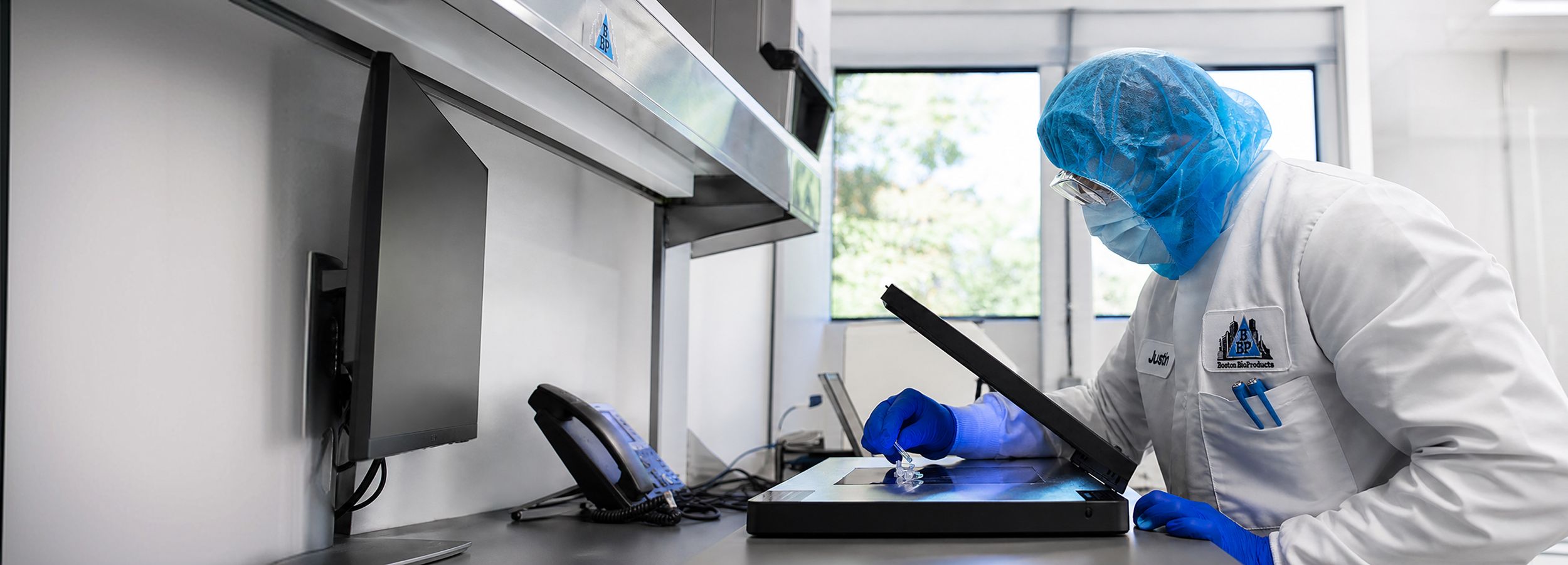
Researcher performing RNase testing in a molecular biology lab

RNase Testing

RNase is common in living cells and breaks down RNA, which can contaminate reagents if it is not carefully controlled. Learn more about RNase testing for your custom reagent and how it can help protect both your reagents and your data.

Considerations & Limitations

Considerations and Limitations of RNase Testing

Clean Work Area and Equipment

Prioritize the cleanliness of workspaces, pipettes, and incubators by using nuclease decontamination solutions.

Protective Attire

Wear laboratory coats and gloves to prevent particulate materials from contaminating samples. Change gloves after touching potentially contaminated surfaces.

RNase-Free Materials

Use tips, tubes, and equipment known to be RNase-free to avoid contamination.

Biological Hood Usage

Perform the assay in a biological safety cabinet or a low-traffic area away from air vents or open windows to minimize external contamination.

Adjust pH and Salt Concentration

Ensure that the pH and salt concentration of samples are adjusted to optimal conditions for RNase assay, as these factors can affect enzyme activity.

Sterilization

Autoclave or sterile filter buffer solutions immediately after preparation to prevent contamination.

Inhibitors and Activators

pH of the Buffer Solutions

RNase activity is optimal at neutral pH (i.e., 7 to 8). Hence, it is important that the pH of the samples is adjusted to neutral pH before subjecting them to RNase assay

Ionic Concentration in the Buffer Solutions

Solutions with high ionic strength are known to inhibit RNases. Therefore, it is necessary that the samples with high salt concentrations are diluted (up to 5-10 mM) appropriately before performing the assay

Presence of Divalent Cations in Buffer Solutions

The presence of Ba2+, Co2+, Pb2+, As3+, and Cu2+ cations inhibit RNase activity.

Presence of Chelators in Buffer Solutions

Presence of chelators such as EGTA and EDTA inhibit RNase activity.

Presence of Reducing Agents in Buffer Solutions

The presence of reducing agents such as β-mercaptoethanol, dithiothreitol (DTT), dithioerythritol (DTE), and reduced glutathione (GSH) inhibit the RNase activity.

Presence of Chaotropic Agents

Chaotropic agents such as guanidine thiocyanate, guanidine hydrochloride and urea denature the RNases and thus inhibit their activity.

Gel Loading Buffers or Colored Solutions

Colored solutions such as Protein loading Dye or DNA loading dye solution interfere with DNase assays, In addition to the dyes in these loading solutions the presence of SDS, EDTA, or β-mercaptoethanol further inhibits RNase activity.

DEPC Treatment

While DEPC treatment can inactivate RNases in some solutions, avoid using it in buffers containing primary and secondary/tertiary amines, such as Tris and HEPES, as it may not be effective and could impact buffering capabilities.

References

  1. Kunitz M. (1946). A spectrophotometric method for the measurement of ribonuclease activity. J Biol. Chem.;164(2):563–8.
  2. Lee, P.Y., Costumbrado, J., Hsu, C.Y., Kim, Y.H. (2012). Agarose gel electrophoresis for the separation of DNA fragments. J. Vis. Exp. (62):3923
  3. Findlay, D., Herries, D. G., Mathias, A. P., Rabin, B. R., and Ross, C. A. (1961). The active site and mechanism of action of bovine pancreatic ribonuclease. Nature 190: 781-784.
  4. Gersten, D. M., and Gabrielt, O. (1992). Staining for Enzymatic Activity after Gel Electrophoresis. Anal. Chem. 203: 181-186.
  5. Greiner-Stoeffele T, Grunow M, Hahn U. (1996). A general ribonuclease assay using methylene blue. Anal. Biochem. 240 (1):24-8
  6. Roth, J.S. and Milstein, S.W. (1952). A new assay method with P labeled yeast ribonucleic acid. J. Biol. Chem. 196(2):489-49
  7. Bechhofer, D. A. and Deutscher, M. P. (2019). Bacterial ribonucleases and their roles in RNA metabolism. Crit. Rev. Biochem. Mol. Biol.54(3): 242–300.
  8. Sato, S and Takenaka, S (2014). Highly Sensitive Nuclease Assays Based on Chemically Modified DNA or RNA. Sensors (Basel). 14(7): 12437–12450.
  9. Shapira, R. (1962). A spectrophotometric method for the measurement of ribonuclease activity. Analytical Biochemistry, 3, 308-320.
  10. Wagner, A.P., Lieselotte, M. C., and Wagner. P., (1983). A simple spectrophotometric method for the measurement of ribonuclease activity in biological fluids. J Biochem. Biophys. Methods. 8(4):291-297.
Boston BioProductsBoston BioProducts
Your quote is empty.
My Cart
  • Products
    Product
    • Buffers
    • Solutions
    • Media
    • Chemical Powders
    • Water
    • Bioprocessing
    • Custom Buffer Manufacturing
    Buffers
    • Good's Buffers
    • Buffered Saline
    • Electrophoresis Buffers
    • Lysis Buffers
    • Standard Buffers
    • Hybridization Buffers
    • HPLC/FPLC Buffers
    • qPCR Buffers
    View all Buffers
    Solutions
    • Acid & Base
    • Balanced Salts
    • Metal
    • EDTA & EGTA
    • Antibiotics
    • Detergent
    • Acrylamide
    • Sugar
    • Fixative
    • Inhibitor Solutions
    • Enzyme
    • Dyes & Stains
    • Luminol & Oxidizing Agents
    View all Solutions

    Featured Products

    Phosphate Buffered Saline, PBS (pH 7.4, Sterile-Filtered)

    Phosphate Buffered Saline, PBS (pH 7.4, Sterile-Filtered)

    Phosphate Buffered Saline (PBS) is a versatile buffer solution widely used in biological research due to its isotonic and non-toxic nature to cells. With a pH range of 7.0 to 7.4, PBS mimics the osmolarity and ion concentrations of the human body, making it ideal for a variety of applications including protein purification, as wash buffer in Western blot and immunoassays, sample dilution, and as a transport medium for tissue samples as well as RNA viruses.

    View product
    Tris-Glycine-SDS Running Buffer (10X)

    Tris-Glycine-SDS Running Buffer (10X)

    The Tris-Glycine-SDS Running Buffer is the most commonly used buffer for the separation/resolution of proteins in their denatured state during SDS-PAGE. It serves as both the anode and cathode buffer during gel electrophoresis and helps to regulate the pH of the electrophoresis system. When diluted to 1X, this buffer regulates pH levels at a pH of about 8.6, which provides reproducible separation of a wide range of proteins into well-resolved bands, depending upon the gel percentage used.

    View Product
    Transfer Buffer (10X, Electro Blotting)

    Transfer Buffer (10X, Electro Blotting)

    The 10X Transfer Buffer-Electro Blotting is the most common buffer used in the wet transfer system for the electro transfer of proteins. The buffer facilitates protein transfer from SDS-PAGE gels to nitrocellulose or PVDF membranes. Ready-to-use solution once diluted to 1X working concentration; Methanol can be added to the final concentration of 10-20% (v/v) as per user protocol. This buffer prevents the gel from swelling during transfer and enhances efficiency of low abundancy proteins and protein binding to nitrocellulose membrane. This buffer is also used as a running buffer for native protein gel electrophoresis.

    View Product
  • Custom
    • Custom Buffer Manufacturing
    • GMP Reagent Manufacturing
    • White-Label and Kitting
    • Custom Buffer Builder
  • Services
    • Reagent Stability Studies
    • Quality Control Testing
    • QC Test Builder
  • Quality
    • Quality Assurance
    • Our Facilities
    • Quality Control Testing
      • Absorbance Testing
      • Bioburden Testing
      • Conductivity Testing
      • Density Testing
      • DNase Testing
      • Endotoxin Testing
      • Microbial Growth Testing
      • Osmolality Testing
      • pH Testing
      • Protease Testing
      • Refractive Index Testing
      • RNase Testing
      • Stability Testing
      • Sterility Testing
      • Viscosity Testing
      • Bubble Point (BP) Filter Integrity Testing
    • Documentation Request
  • Resources
    • Content Hub
      • Product Literature
      • Safety Data Sheets
      • Publications
      • Case Studies & Infographics
      • Protocols
      • Articles & Insights
    • Buffer Calculator
    • Applications
      • Molecular Biology
        • FPLC Protein Purification
        • SDS PAGE
        • HPLC Buffers
        • Plasmid Isolation
        • ELISA Buffers
        • Blood Lysis Buffers
      • Cell Culture
        • Cell Culture Buffers
        • Yeast and Bacteria Culture Media
        • Western Blotting Buffer
        • IHC Buffer
      • Diagnostics
        • Immuno Diagnostics
      • Bioproduction
        • Upstream Bioprocessing
        • Downstream Bioprocessing
    • FAQs
    • Order Support
  • Company
    • About Us
    • Contact Us
    • Careers
    • News
    • Events
My AccountContact Us
Your cart is empty.