Introduction
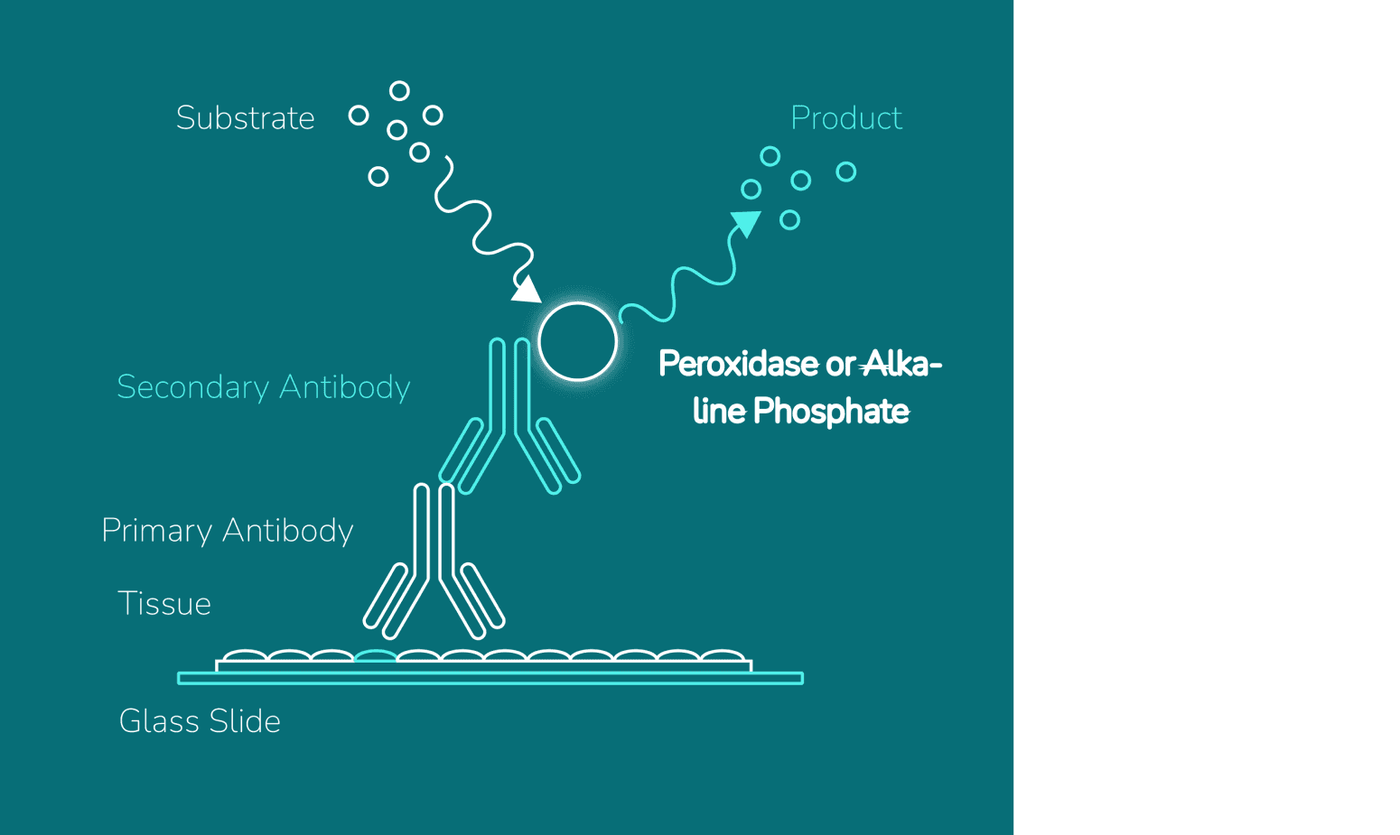
Immunohistochemistry (IHC) detects and visualizes antigens in tissue sections using antibodies. It preserves the spatial context of proteins within tissues and enables researchers to study cellular localization, tissue architecture, and disease pathology in detail. This makes IHC a valuable tool in cancer research, neuroscience, and pathology.
Poor fixation, high background noise, or weak staining can compromise data integrity and reproducibility. Using high-quality reagents, such as those from Boston BioProducts, in combination with an optimized IHC protocol helps ensure strong, specific staining with minimal background interference.
Read on for a clear IHC step-by-step guide, troubleshooting tips, and best practices to help you confidently achieve precise, high-quality results.